
Breast cancer
Breast cancer is the most common cancer in women. Early detection and modern treatments have significantly improved survival rates. At WEGE Klinik, care is guided by precision medicine and delivered with attention to your individual needs.
Patients bring their medical reports and imaging for an in-depth consultation, where treatment options, timing, and possible side effects are discussed in detail. Patients usually present with a recommendation from a multisiciplinary tumour board, sometimes from their gynecologist or for a second opinion.
Our treatments are guided by innovation and delivered with deep human care. Experience precision medicine in a place where you are truly seen and supported.
*Mandatory fields
Specialist in breast care
Led by Prof. Dr. Michael Pinkawa with over 20 years of experience, radiotherapy and brachytherapie at WEGE Klinik is performed by an interdisciplinary team, combining technical precision and compassionate care. Our team
Treatments at WEGE
We use high-precision radiotherapy technology to maximize effectiveness and reduce side effects in the breast cancer treatments. Treatment decisions are made by a multidisciplinary and highly experienced team. We care for patients after breast-conserving surgery as well as those who require mastectomy. Our goal is to achieve optimal tumour control with minimal impact on healthy tissue. Our approach includes external beam radiotherapy, respiratory control as DIBH (deep inspiration breath control), and brachytherapy, depending on the patient’s needs.
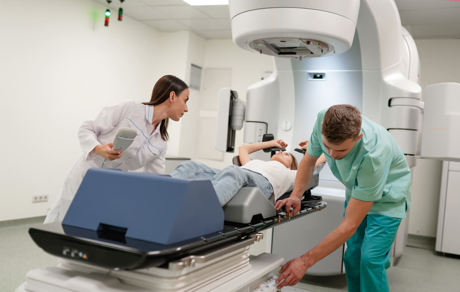
External beam radiotherapy (EBRT) and deep inspiration breath hold (DIBH) technique
Postoperative external beam radiotherapy is typically delivered over 3 weeks (15 sessions).
Postoperative external beam radiotherapy is usually performed over 15, fractions, thus over a period of 3 weeks with 5 weekly fractions. For more favourable tumours, a partial breast irradiation might be the best method. For tumours with specific risk factors, the dose to the initial tumour site is increased in comparison to the doce to the whole breast (designated as “boost”).
The exact detection of the patient surface and adaptation to the breathing phases is essential for an optimal treatment, including the best possible dose coverage of the prescibed dose in the target (breast or chest wall, possibly lymph node areas) and the best posible protection of healthy tissue outside the target volume. Radiotherapy of the left breast requires a treatment only in deep inspiration. In deep inspiration, the lung volume expands and increases the distance of the breast tissue to the heart. Thus, radiotherapy in deep inspiration is associated with the best possible heart protection.
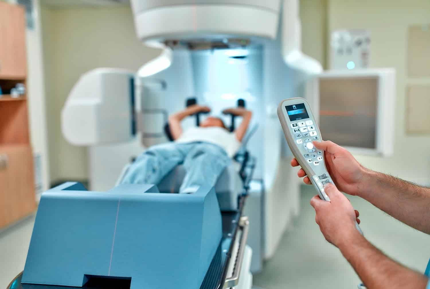
Patients in WEGE are treated with the most sophisticated system for surface detection and deep inspiration breath hold technique (ExacTrac Dynamic, Brainlab). The system combines the detection of the surface with the termal surface information. Baseline imaging information for planning are acquired during an appointment for treatment planning. Computed tomography images are acquired in normal breathing and in deep inspiration breath hold. With the ExacTrac Dynamic system, no addition skin markings are needed for patient positioning.
The 4D Thermal Camera creates a highly accurate and reliable hybrid thermal surface by correlating the patient’s heat signature to their reconstructed 3D surface structure. To achieve this, 300.000 3D surface points are acquired and matched to the heat signal generated by the thermal camera, creating another dimension to track their position. The patient receives a continuous visual feedback of the required surface position. Radiation is only triggered in the correct phase of deep inspiration. Thus, depending on the duration of the breath hold, each patient determines the duration of the radiation phases.
Adjuvant breast radiotherapy
Most breast cancer patients receive radiotherapy after breast-conserving surgery. This approach provides similar tumour control and survival to mastectomy in early-stage cases.
The majority of patients with the diagnosis of breast cancer receive radiotherapy of the breast after breast conserving operation. The local cancer excision in combination with radiotherapy has shown to be associated with the same tumour control and survival as the removal of the complete breast (mastectomy). Only in patients with more advanced, larger tumours or tumours with an invasion of the skin or chest wall, the removal of the complete breast is still recommended. In these patients, breast reconstruction methods are discussed and postoperative radiotherapy of the remaining chest wall is decided after discusion of individual risk factors in a multidisciplinary tumour conference in the respective breast cancer centre.
Apart from the radiotherapy of the breast or chest wall, the decision of treatment of locoregional lymph nodes made. Locoregional lymph nodes include axillary, infra- and supraclavicular lymph nodes (lateral lymph nodes in the direction of the axilla and neck) and internal mammary lymph nodes (medial lymph nodes near the sternum). Patients with positive lymph nodes usually need a postoperative radiotherapy of locoregional lymph nodes. Radiotherapy of the lymph nodes might be recommended instead of an operation, as side effects of radiotherapy have been shown to be considerably lower in comparison to an operation (lymph oedema) in recent studies without compromising tumour control.
Patients present at our clinic for their first appointment with their reports, prior images and multidisciplinary tumour conference recommendation. The treatment is discussed and explained in the context of the individual patient situation, including the timing, duration, potential problems that might occur and individual questions.
Brachytherapy
Brachytherapy involves placing a radioactive source directly into the breast tissue, allowing very focused partial breats treatment and minimal exposure to nearby organs. Doses are higher per session, so treatment is shorter than with external radiotherapy.
Brachytherapy is based on the placement of a radioactive source in the target tissue. In contrast to external beam radiotherapy, the dose is not applied from the outside through the skin and potentially other organs, but directly placed in the target tissue. The consequence is the best possible treatment conformality (best focus on a specific area) and the lowest doses to organs as lung or heart. Fraction doses in brachytherapy are usually considerably higher and treatment duration considerably shorter in comparison to external beam radiotherapy.
Adjuvant breast cancer brachytherapy is a well established treatment that has been evaluated in several randomized studies with many thousands of patients, thus endorsed by all national and international guidelines for breats cancer treatment. An important advantage is an improved cosmesis, including reduced long-term impact on the skin and breast tissue. A disadvantage is the invasive character of the treatment. Brachytherapy is always a partial breats irradiation, recommended only for patients with smaller, less aggressive tumours without lymph node involvement.
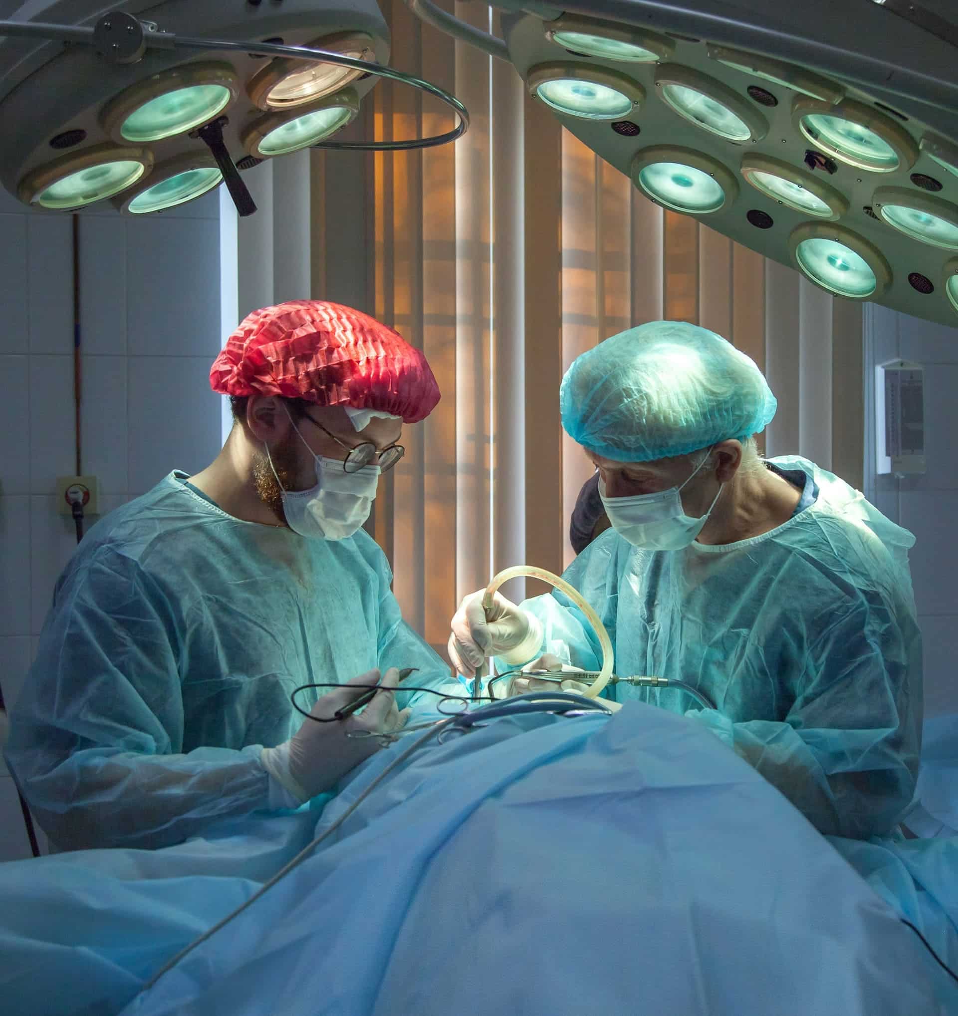
Adjuvant breast cancer brachytherapy commonly applied in 8 twice daily fractions during an inpatient stay at WEGE Klinik over a period of 5 days (usualy Monday to Friday). At the first day, applicator catheters are implanted in the breast under general anaesthesia. With the implanted catheters in situ, a planning computed tomography is performed as the basis for three-dimensional treament planning. The first brachytherapy fraction is commonly applied on the afternoon of the first day. During treatment, the radioactive source (Iridium-192) is placed in the breast tissue via the applicator catheters. The source remains for a specific time at specific places. These times and places are calculated in the treatment plan to deliver the required dose distribution. The duration of each fraction is about 15 minutes.
What is breast cancer? |
Anatomy and function of the breast |
How breast cancer develops |
Symptoms and risk factors |
|---|---|---|---|
|
Breast cancer is a disease in which cells in the breast grow uncontrollably, forming a tumour. If untreated, it can invade nearby tissues or spread to other parts of the body, becoming life-threatening.
Most breast cancers begin in the milk ducts (ductal carcinoma) or lobules (lobular carcinoma), which are responsible for milk production. Early detection is key, as localized tumors are easier to treat and less likely to spread through the lymphatic or blood systems. |
The breast contains lobules (which produce milk), ducts (which carry it to the nipple), and fatty and connective tissue (which give shape and support). Blood vessels nourish the breast, and lymphatic vessels help with immune defense.
Hormones like estrogen and progesterone influence breast development and function. Knowing breast anatomy helps explain how most cancers start—in the ducts or lobules—and how they can spread through lymph nodes or blood circulation. |
Breast cancer begins when DNA damage causes certain breast cells to grow and divide uncontrollably. These cells form a tumour that can stay localized or spread to nearby or distant areas.
Some tumours grow in response to hormones like estrogen or progesterone, or due to overproduction of the HER2 protein. The cancer may spread via the lymphatic system or bloodstream, which is why early detection and personalized treatment are so important. |
Common symptoms include a lump in the breast or armpit, changes in breast shape or skin, nipple discharge or inversion, and persistent pain. While not all changes mean cancer, they should be evaluated.
Risk factors include age, family history, BRCA1/BRCA2 mutations, early menstruation or late menopause, hormone therapy, obesity, alcohol use, and lack of physical activity. Regular screening can help detect cancer before symptoms appear. |
Standard treatment typically lasts about 3 weeks, with one session per weekday. Some patients may qualify for shorter courses or partial breast irradiation, depending on their case.
No, the treatment itself is painless. You lie still while the machine delivers radiation. Some skin irritation or fatigue may appear later, but the sessions are not physically uncomfortable.
Yes. At WEGE, we use advanced techniques like deep inspiration breath hold (DIBH) to increase the distance between the heart and the breast during treatment. This helps protect the heart and reduces the risk of long-term cardiac side effects.
Many women continue to work or care for loved ones, though you may need to adjust your routine. It’s okay to ask for help — treatment affects everyone differently.
Testimonials
At WEGE, we are committed to supporting our patients and their families throughout the breast cancer journey, offering expert care and understanding every step of the way.
 Maria S., 60 – Bonn, Germany
“From the moment I walked into WEGE Klinik, I knew I was in good hands. The multidisciplinary team worked seamlessly to provide a treatment plan tailored to my needs. Their dedication and expertise were evident every step of the way.”
Sarah K., 55 – Munich, Germany
“The medical team at WEGE Klinik is truly outstanding. Their expertise, clarity, and genuine care made all the difference during my treatment. I felt safe, respected, and fully supported. I highly recommend them to anyone facing breast cancer.”
If you have general questions, you can contact us by phone or e-mail. We will then get in touch with you as soon as possible.
WEGE Klinik:
Phone: +49 228 5306 0
Email: info@wegeklinik.com
Web: www.wegeklinik.com
WEGE MVZ:
Phone: +49 228 5306 202
Email: info@wegemvz.com
Web: www.wegemvz.com