
Gynaecological tumours
At WEGE Klinik, we care for patients with gynaecological tumours using highly precise radiotherapy tailored to the anatomy and condition of each person. Our focus is on effective, individualised treatment that protects healthy tissue while targeting the tumour with accuracy.
Patients are often referred by their gynaecologist or tumour board for a second opinion or radiotherapy planning. During the first appointment, our team reviews all previous imaging and reports to assess the best strategy. We explain the type of radiotherapy, timing, and possible side effects, and address all patient concerns in a clear and supportive way.
Our treatments are guided by innovation and delivered with deeply human care. Experience precision medicine in a place where you are truly seen and supported.
*Mandatory fields
Specialists in gynaecological tumours care
Led by Prof. Dr. Michael Pinkawa, our radiotherapy team has extensive experience in treating pelvic gynaecological tumours. Radiotherapy at WEGE Klinik is performed by an interdisciplinary team that brings together clinical expertise and personalised care.
Meet our team
Treatments at WEGE
The appropriate radiotherapy treatments for gynaecological tumours depend on the type, stage, and location of the tumour, as well as whether the goal is curative or adjuvant. At WEGE Klinik, treatment is tailored to each patient using state-of-the-art technology and imaging.
Gynecological tumours (pelvis)
Gynecological tumours in the pelvis include tumour of the uterus (corpus and cervix), the vagina and vulva. Tumours of the vagina are rarely occurring tumours with an important role for radiotherapy and brachytherapy. Ovarian tumours are commonly treated with radiotherapy only in a metastatic situation.
Radiotherapy is applied as single modality or in combination with a systemic therapy (as chemotherapy and immunotherapy) as definitive treatment (without an operation) or postoperative treatment in case of specific risk factors that require an additional treatment and offer the best possible chances to cure the disease.
Uterine corpus tumour
For the majority of uterine corpus tumours, the resection of the uterus and often also locoregional pelvic lymph nodes is the initial treatment after diagnosis. The decision for postoperative treatment is based on individual factors, including the tumour stage (considering the size of the tumour, invasion of neighbouring tissues or lymph nodes) and specific pathologically determined factors (immune histochemistry and molecular genetics). For low-risk tumours, no postoperative treatment is needed. Patients usually present in WEGE MVZ for the first appointment after recommendation for a specific treatment in a multidisciplinary tumour board. Prior reports and imaging is needed for a detailed assessment and discussion in our MVZ.
For intermediate risk tumours, vaginal brachytherapy as the only modality is frequently recommended. This is an outpatient treatment that is usually performed in five fractions, with a single fraction per week. For vaginal brachytherapy, an applicator is inserted into the vagina, similar to an intravaginal ultrasound probe. After CT-based three-dimensional treatment planning, a radioactive Iridium-192 source is transferred into the applicator. It remains for a few seconds at precalcualted positions. The treatment duration takes only 3-5 minutes. The patient does not feel any effect during this treatment.
In a high-risk tumour, the risk or tumour spread into pelvic nodes or areas outside the pelvis is increased. Therefore, treatment commonly includes a chemotherapy and an external beam radiotherapy. An external beam radiotherapy implicates the treatment of the vaginal vault and pelvic nodes. The treatment is performed in daily fractions, five days a week, over about six weeks. External beam radiotherapy is applied as a so called volumetric modulated arc therapy (VMAT), delivering the dose during continuous rotation of linear accelerator gantry around the patient and continuous movement of the leafes that are forming the beam from the respective direction.
Cervical tumour
Smaller cervical tumours are rected locally (cone biopsy) or resection of the uterus with pelvic lymph nodes. Depending on individual factors, as tumours size or with tumour spread into pelvic nodes, an additional might be needed. This treatment often includes a combination of external beam radiotherapy, as described above, with a simultaneous chemotherapy.
Gynecological brachytherapy
Standard treatment for tumours extending outside the cervix or uterus or with extension into pelvic nodes is a combined radio chemotherapy (systemic treatment might also include an immunotherapy) and an additional brachytherapy.
Brachytherapy is based on the placement of a radioactive source (Iridium-192) in the target tissue. In contrast to external beam radiotherapy, the dose is not applied from the outside through the skin and potentially other organs but directly placed in the target tissue. The consequence is the best possible treatment conformality (best focus on a specific area) and the lowest doses to organs as bladder or bowel. Fraction doses in brachytherapy are usually considerably higher and treatment duration considerably shorter in comparison to external beam radiotherapy.
Brachytherapy is an essential part of cervical cancer treatment. It is only performed in specific specialized centres, as specific expertise is needed for this treatment. Three-dimensional treatment planning is based on a recent MRI and a planning CT with the applicator in place. The applicator is placed intravaginally and into the cervical canal. Commonly, the placement is performed under anesthesia using applicator needles that are placed into the cervical tissue (interstitial needles). Additional placement of the radioactive source into these needles improves the tumour coverage considerably. It is essential to reach an adequate plan quality, as brachytherapy is a crucial part of the multimodal treatment. In most cases, two fractions are applied with the same applicator in place on two consecutive days and additional two fractions in the following week with a second applicator placement. Interstitial brachytherapy is performed during a short inpatient stay at WEGE Klinik.
Vulvar tumour
An operative resection is the first step in the treatment of vulvar tumours. If resection margins are not adequate or tumour cells detected in lymph nodes (usually inguinal lymph nodes), a postoperative radiotherapy (sometimes radio chemotherapy, radiotherapy combined with chemotherapy) will be often recommended in the multidisciplinary tumour board. The target of the external beam radiotherapy can be the vulva and/or lymph node areas, depending on the individual risk.
If resection is not possible, a primary radiotherapy or radio chemotherapy of the vulvar tumour and locoregional lymph node regions is performed. This is usually an external beam radiotherapy in 30-35 daily fractions, five fractions per week. In selected cases, brachytherapy is applied as a boost to external beam radiotherapy (boost = increased dose in the high-risk macroscopic tumour to increase efficiency). Applicator catheters are implanted in the vulvar tumour under general anesthesia. With the implanted catheters in situ, a planning ultrasound or computed tomography is performed as the basis for three-dimensional treatment planning. During treatment, the radioactive source (Iridium-192) is placed in the vulvar tumour via the applicator catheters. The source remains for a specific time at specific places. These times and places are calculated in the treatment plan to deliver the required dose distribution. The duration of each fraction is about 5-10 minutes. The number of fractions is depending on the individual case (tumour volume, tumour expansion, neighboring tissues/organs).
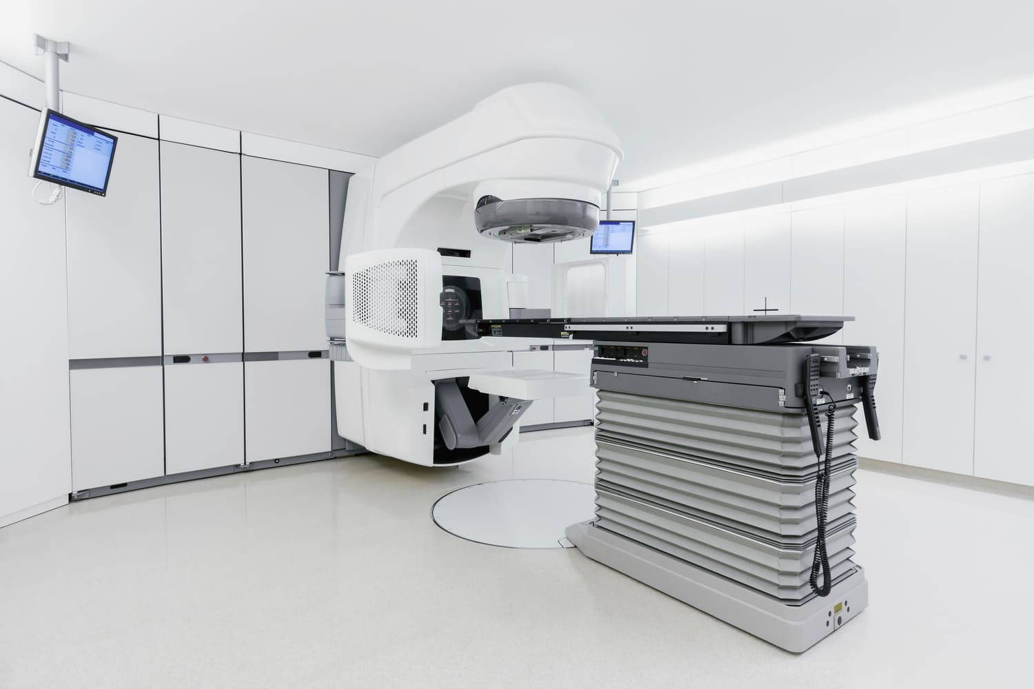
External beam radiotherapy (EBRT)
External beam radiotherapy is used to treat many gynaecological tumours, particularly after surgery or when surgery is not possible. It involves directing radiation at the tumour area from outside the body.
External beam radiotherapy is used to treat many gynaecological tumours, particularly after surgery or when surgery is not possible. It involves directing radiation at the tumour area from outside the body.
EBRT is a core approach, often used after surgery or as primary treatment in cervical, endometrial, and vulvar cancers. It is also indicated when pelvic lymph nodes are involved.
At WEGE, EBRT is delivered using precision-guided systems such as the Elekta Versa HD, and every plan begins with advanced 3D imaging, including spectral CT scanning for optimal anatomical accuracy.
Treatments are typically delivered over several weeks in daily sessions. The goal is to reduce recurrence and control the disease while maintaining organ function.
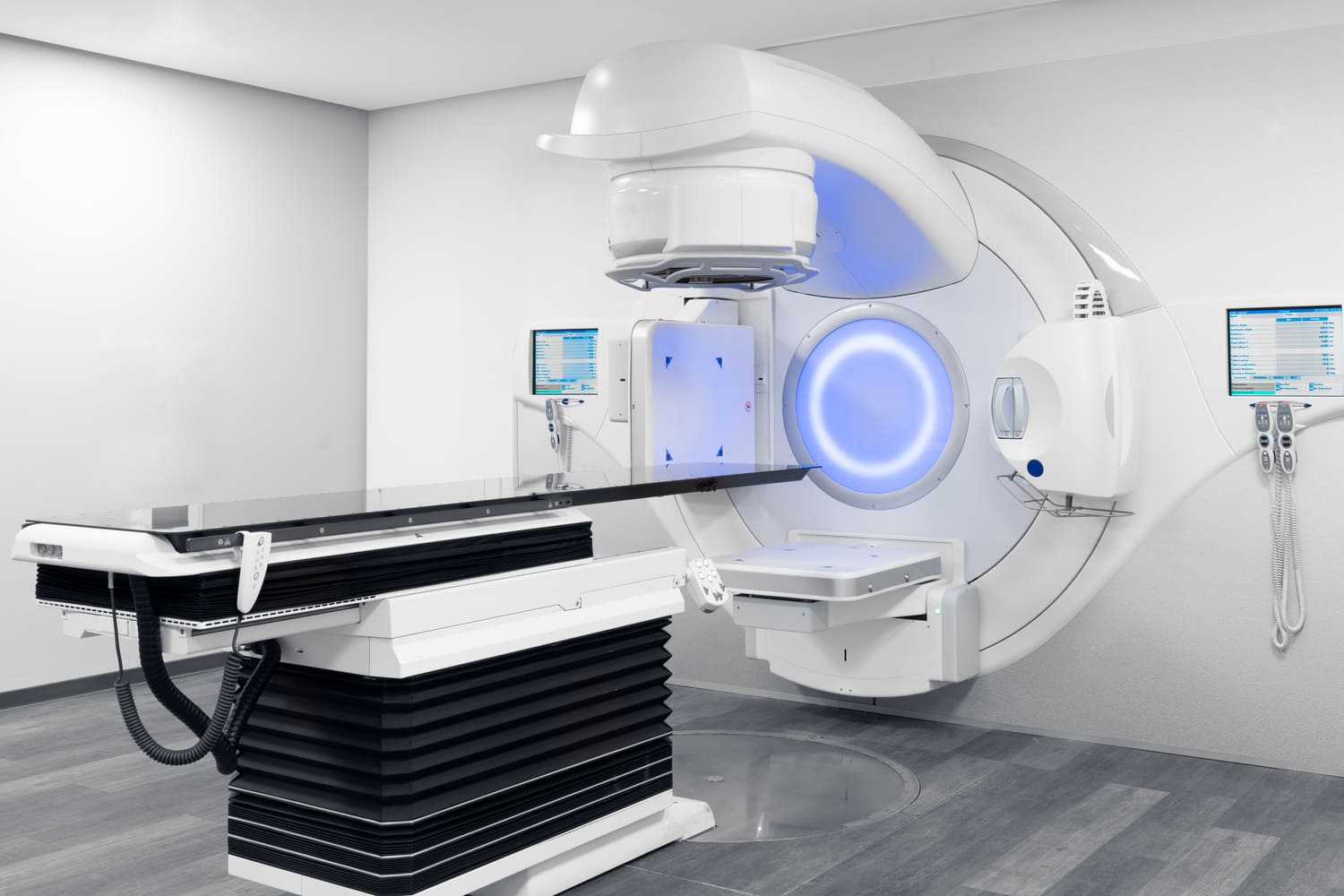
Volume-modulated arc therapy (VMAT)
It allows the radiation dose to be shaped to the tumour volume while reducing exposure to nearby organs such as the bladder, rectum, and small intestine. It is especially indicated for complex pelvic tumours, cervical cancer with lymph node involvement, cases with a high risk of rectal or bladder toxicity, and conservative treatment in frail patients.
VMAT enables efficient and highly conformal dose delivery. The technology adjusts the beam continuously as it rotates around the patient, making sessions faster and more comfortable. It is an excellent option for treating irregular areas or multiple targets, such as the uterus and lymph nodes.
Stereotactic radiotherapy with ExacTrac Dynamic
It delivers high doses in a small number of sessions with sub-millimetre precision, requiring real-time image-guidance systems. It is indicated for isolated pelvic lymph node metastases, small inoperable recurrences, and patients who cannot tolerate long treatment courses.
It delivers high doses in a small number of sessions with sub-millimetre precision, requiring real-time image-guidance systems. It is indicated for isolated pelvic lymph node metastases, small inoperable recurrences, and patients who cannot tolerate long treatment courses.
At WEGE Klinik, this is performed using the Brainlab ExacTrac Dynamic system in combination with the Elekta Versa HD linear accelerator. This technology enables real-time monitoring of patient positioning and breathing, ensuring sub-millimeter accuracy and safe delivery of high-dose radiation in a small number of sessions.
Brachytherapy
Brachytherapy is an internal radiotherapy technique that places a radioactive source directly inside or near the tumour. It is often used for cervical or endometrial cancer, allowing a high dose to the tumour with minimal exposure to healthy tissue.
Brachytherapy is an internal radiotherapy technique that places a radioactive source directly inside or near the tumour. It is often used for cervical or endometrial cancer, allowing a high dose to the tumour with minimal exposure to healthy tissue.
Brachytherapy is typically performed in a short treatment cycle, often in combination with EBRT. Planning is based on 3D imaging to ensure precise positioning and dose distribution.
What is gynaecological cancer? |
Anatomy and function of the female pelvis |
How gynaecological cancer develops |
Symptoms and risk factor |
|---|---|---|---|
|
Gynaecological cancer refers to malignant tumours that begin in female pelvic organs, including the cervix, uterus, ovaries, vulva, and vagina. Each type has distinct characteristics, but they are all located within the pelvic area and may require radiotherapy as part of treatment. |
The female reproductive system includes the ovaries (which produce eggs and hormones), the uterus (where pregnancy occurs), the cervix (connecting uterus and vagina), and the vulva (external genitalia). These organs are functionally interconnected and located close to the bladder and bowel, which makes precision in radiotherapy especially important. |
Gynaecological tumours begin when normal cells in these organs start to grow uncontrollably. This can be triggered by genetic mutations, infections such as HPV, or hormonal imbalances. Over time, these tumours may invade nearby tissue or spread through the lymphatic system or bloodstream. |
Risk factors include persistent HPV infection (especially for cervical and vulvar cancer), a family history of ovarian, breast, or uterine cancer, obesity, smoking, hormonal imbalance or unopposed oestrogen, and a weakened immune system. |
It depends on the type of tumour and treatment. At WEGE Klinik, we discuss fertility before starting any therapy. Options like egg or embryo preservation may be considered.
Progress is monitored through regular imaging and consultations. We explain all results in detail and are available to answer questions at any point.
Our care team includes professionals in psychological support, as well as access to counselling and peer support groups. We are here to help you through every step.
Testimonials
At WEGE Klinik, we are committed to supporting our patients and their families throughout the gynaecological tumors journey, offering expert care and understanding every step of the way.
 Anneliese, 59 – Münster, Germany
When I was told I had ovarian cancer, I was scared. It’s hard to accept that your life changes in that moment. The professionalism and empathy of the team at WEGE have helped me move forward. I’m deeply grateful to all of you.
Maren, 52 – Düsseldorf, Germany
I was diagnosed with cervical cancer and didn’t know what to expect. At WEGE Klinik, I found a team that gave me both excellent treatment and true reassurance. The radiotherapy was precise, but it was the people who made the difference.
Heike, 60 – Bonn, Germany
I had a recurrence in the pelvic area and was told I couldn’t have surgery again. The stereotactic radiotherapy at WEGE Klinik was my best option. It was short, accurate, and easier than I imagined.
Eva, 57 – Köln, Germany
If you have general questions, you can contact us by phone or e-mail. We will then get in touch with you as soon as possible.
WEGE Klinik:
Phone: +49 228 5306 0
Email: info@wegeklinik.com
Web: www.wegeklinik.com
WEGE MVZ:
Phone: +49 228 5306 202
Email: info@wegemvz.com
Web: www.wegemvz.com
